
Diagnostics
Diagnosis and Early Detection of Keratoconus

Distorted vision and fluctuating visual acuity may be signs of a loss of elasticity in the cornea, which leads to deformation. This can be caused by a rare condition called keratoconus. Thanks to state-of-the-art technology, we are able to diagnose keratoconus at an early stage.
Early Detection of Keratoconus
High-Tech Diagnostics
ANTERION® SS-OCT (Heidelberg Engineering)
CIRRUS® 6000 SD-OCT (Zeiss)
Pentacam® HR Version (Oculus)
Corvis® ST (Oculus)
Keratoconus can only be diagnosed with experience and high-tech equipment
Keratoconus often becomes noticeable between the ages of 15 and 20. In its early stages, it masquerades as astigmatism and is therefore often treated incorrectly. Other early signs include fluctuating visual impairment, nearsightedness, and increasing thinning and warping of the cornea. As the condition progresses, it can lead to progressive keratoconus with a clearly visible bulge of the cornea. Even experienced doctors can only assess its initial symptoms using state-of-the-art diagnostic tools.
The latest OCT technology sets entirely new standards in this regard because it enables higher image resolution and deeper penetration into the tissue. Based on this technology, the new STEP classification system was developed.1 It analyzes the following corneal parameters:
ST value (Stroma Thinnest Thickness): the thinnest point of the middle corneal layer (stroma)—an important indicator of stability
EP value (Epithelial Standard Deviation): indicates the degree of variation in the thickness of the outer cell layer (epithelium)—a highly sensitive early warning sign of early-stage keratoconus
At Premium Eyes, we use the ANTERION® SS-OCT and the CIRRUS® 6000 SD-OCT to examine your eyes as part of our keratoconus diagnostic process, in order to provide you with more precise early detection and treatment monitoring for keratoconus.
Dr. Kristin Lena Artz, FEBO
CIRRUS® 6000
SD-OCT Examination Using the CIRRUS® 6000
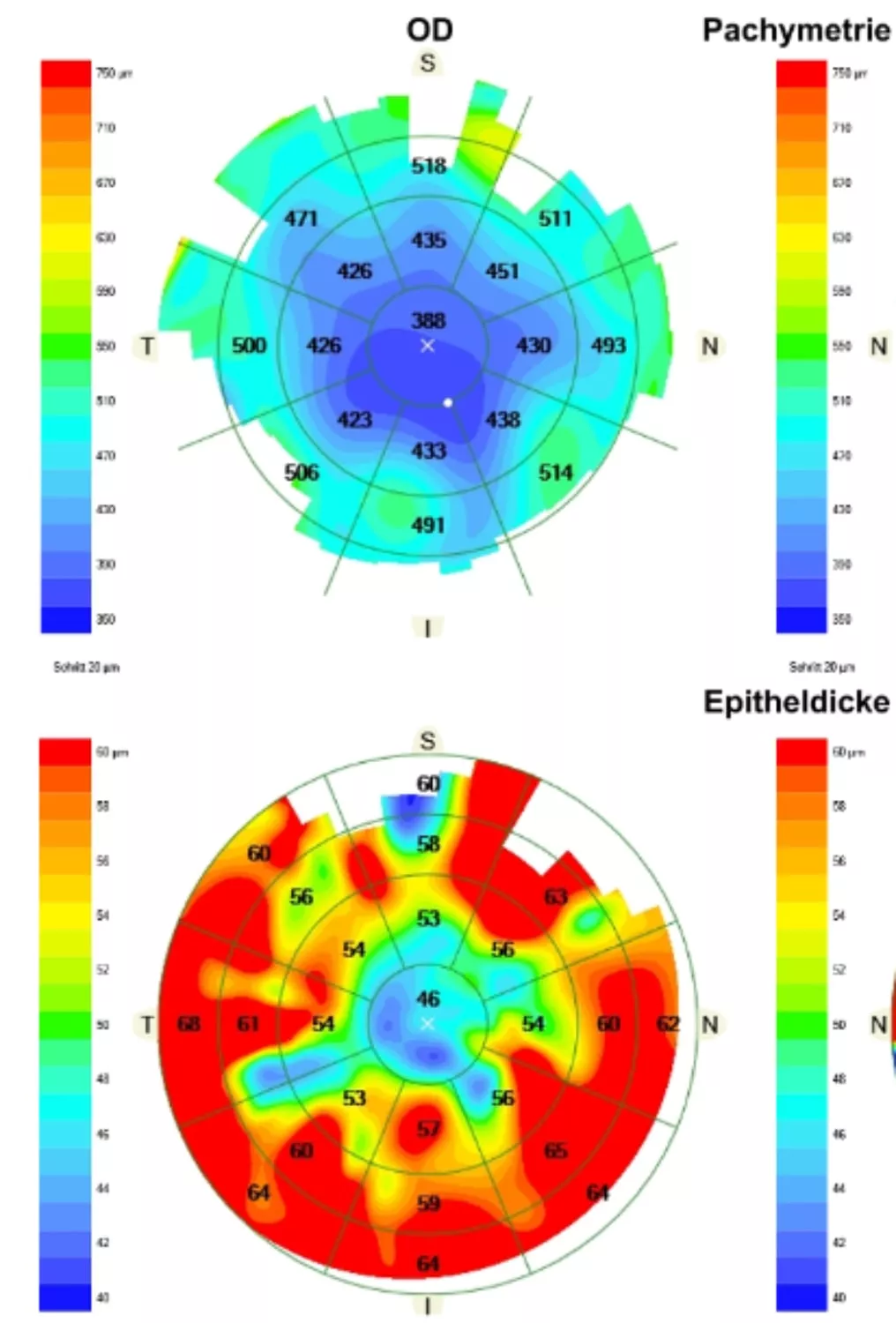
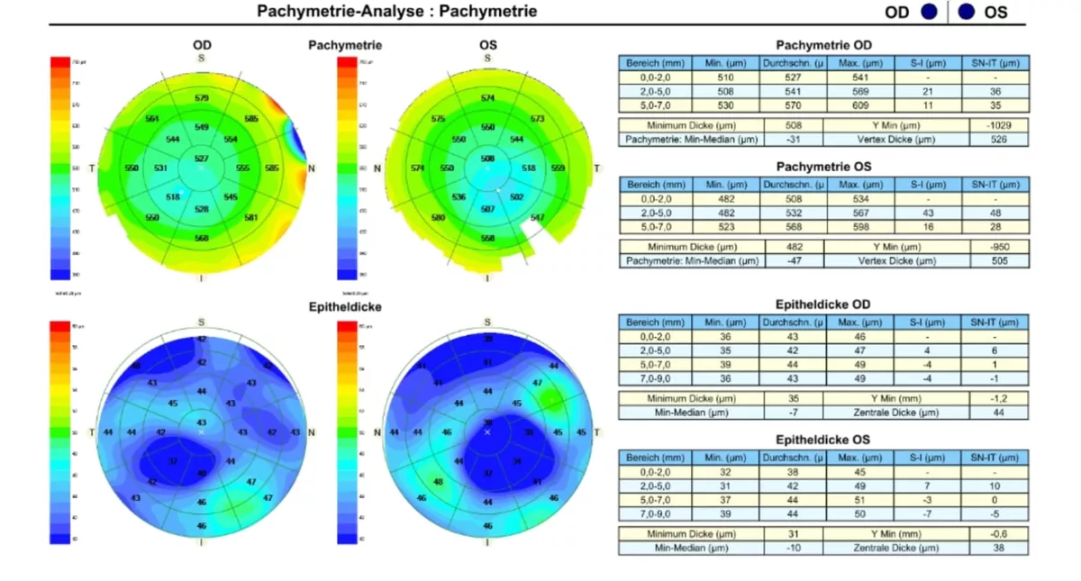
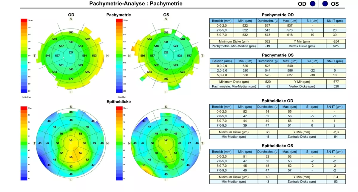
The CIRRUS 6000 (Zeiss), a spectral-domain OCT, or SD-OCT for short, provides, among other things, comprehensive imaging and measurement of the anterior segment for planning and follow-up after refractive procedures, as well as for evaluating the cornea in cases of keratoconus. We primarily use the CIRRUS 6000 to perform an OCT examination and measure the thickness of the corneal epithelium. This measurement directly highlights localized epithelial thinning, which typically occurs in keratoconus. This examination allows for the early classification of keratoconus. In Fig. 1, the areas where the epithelium is already significantly thinned are shown in red in the lower image. Figures 2 and 3, on the other hand, show earlier stages in which the deviations from a healthy eye are even less pronounced.
ANTERION®
ANTERION® Swept-Source OCT
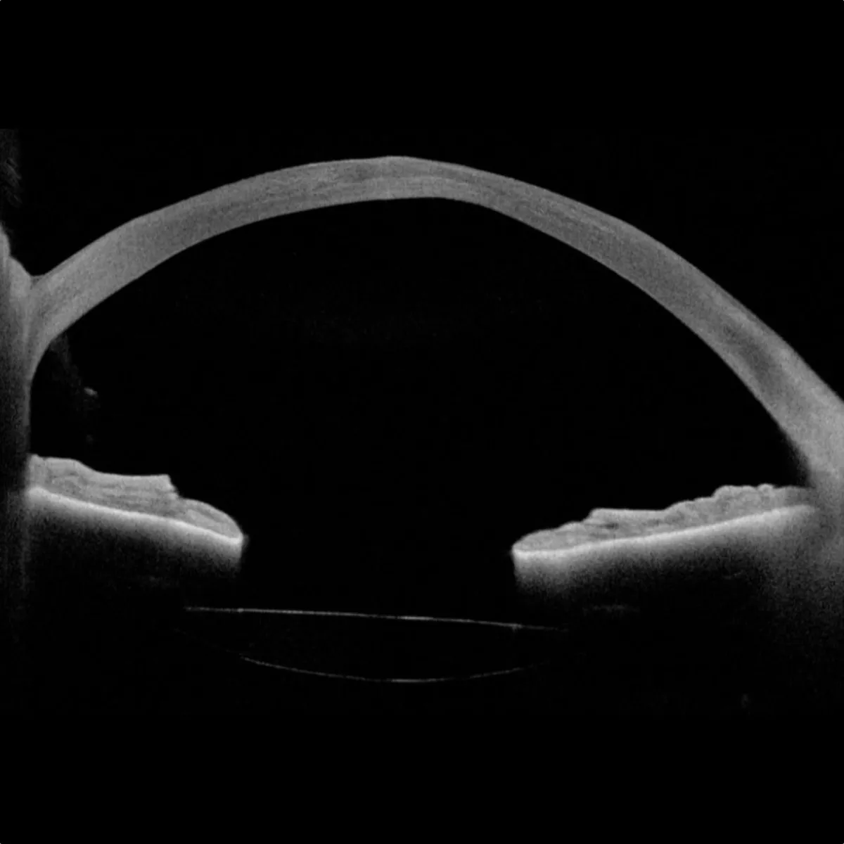
Our keratoconus diagnostics also include an examination using the ANTERION® (Heidelberg Engineering). This device enables the most important examinations and calculations of the anterior segment based on high-resolution swept-source OCT images. In addition to OCT imaging of the cornea, it provides tomographic maps and wavefront analysis. The Anterion combines high-resolution cross-sectional images with precise measurements of all key corneal parameters, including epithelial profile, corneal thickness, corneal curvature, and elevation deviations. As a result, it detects corneal abnormalities at a very early stage and enables classification according to the STEP system. In Fig. 4, you can see the thinning of the cornea quite clearly in the upper region.
Pentacam HR
Diagnostics with the Pentacam® HR
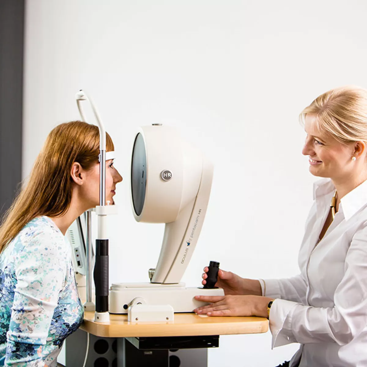
The Pentacam® HR, a state-of-the-art Scheimpflug camera manufactured by Oculus, measures the anterior segment of the eye in just under two seconds. The rotating scan provides numerous measurement points at the center of the cornea. Among other things, the Pentacam examines the shape of the corneal surface (topography) and the posterior corneal surface and performs a full-surface measurement of corneal thickness (pachymetry). It also enables the display of a three-dimensional cross-section of the cornea (tomography) and a 3-D map of the corneal surface.
It visualizes the typical symptoms of keratoconus and offers tomography-based keratoconus detection and classification using the Belin ABCD Keratoconus Staging and the Belin ABCD Progression Display. You can read more about the Belin keratoconus classification on the page about keratoconus.
Corvis® ST
Corvis® ST Non-Contact Tonometer
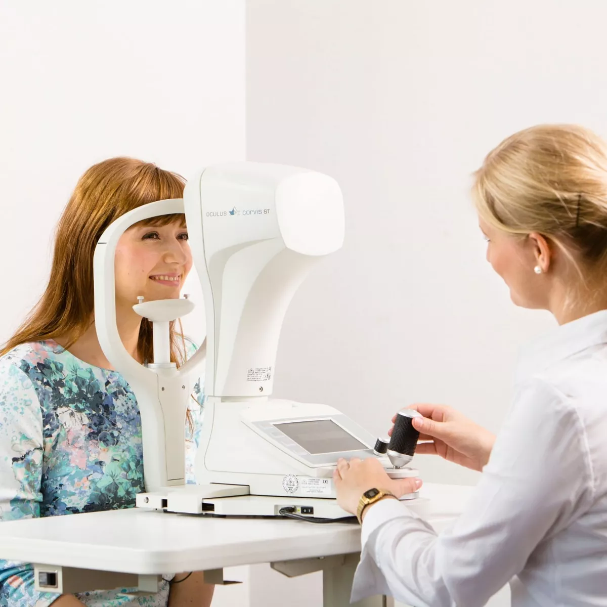
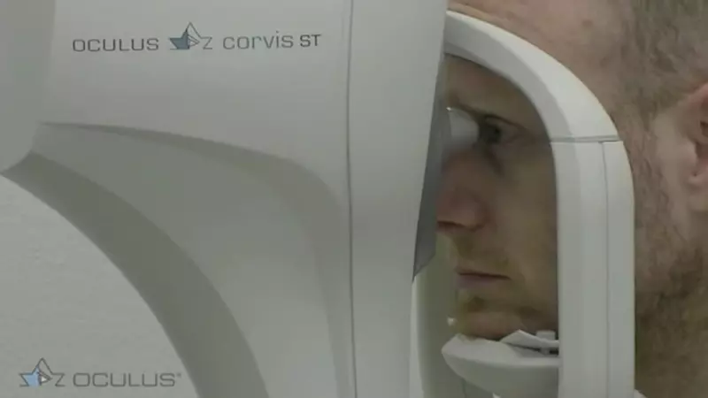
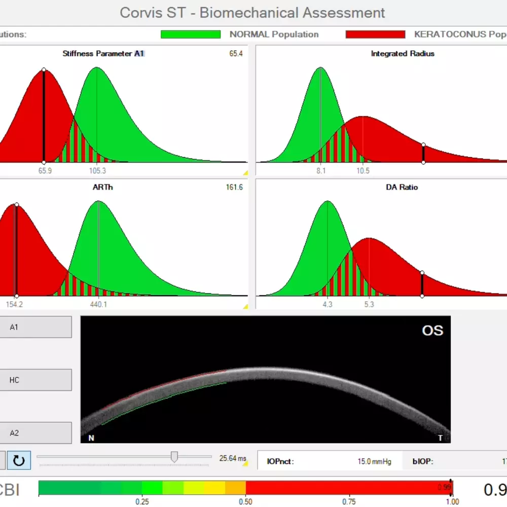
The Oculus Corvis® ST non-contact tonometer has continued to prove its worth in the early detection of keratoconus and for monitoring treatment outcomes following cross-linking: It measures corneal thickness and the cornea’s biomechanical response after it has been exposed to a puff of air. The cornea of an eye with keratoconus exhibits characteristic vibrations that significantly simplify the diagnosis of the disease.
With a Healthy Eye
Healthy Eye
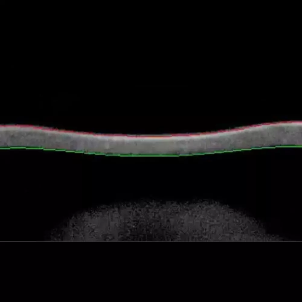
Healthy Eye
Eye with keratoconus
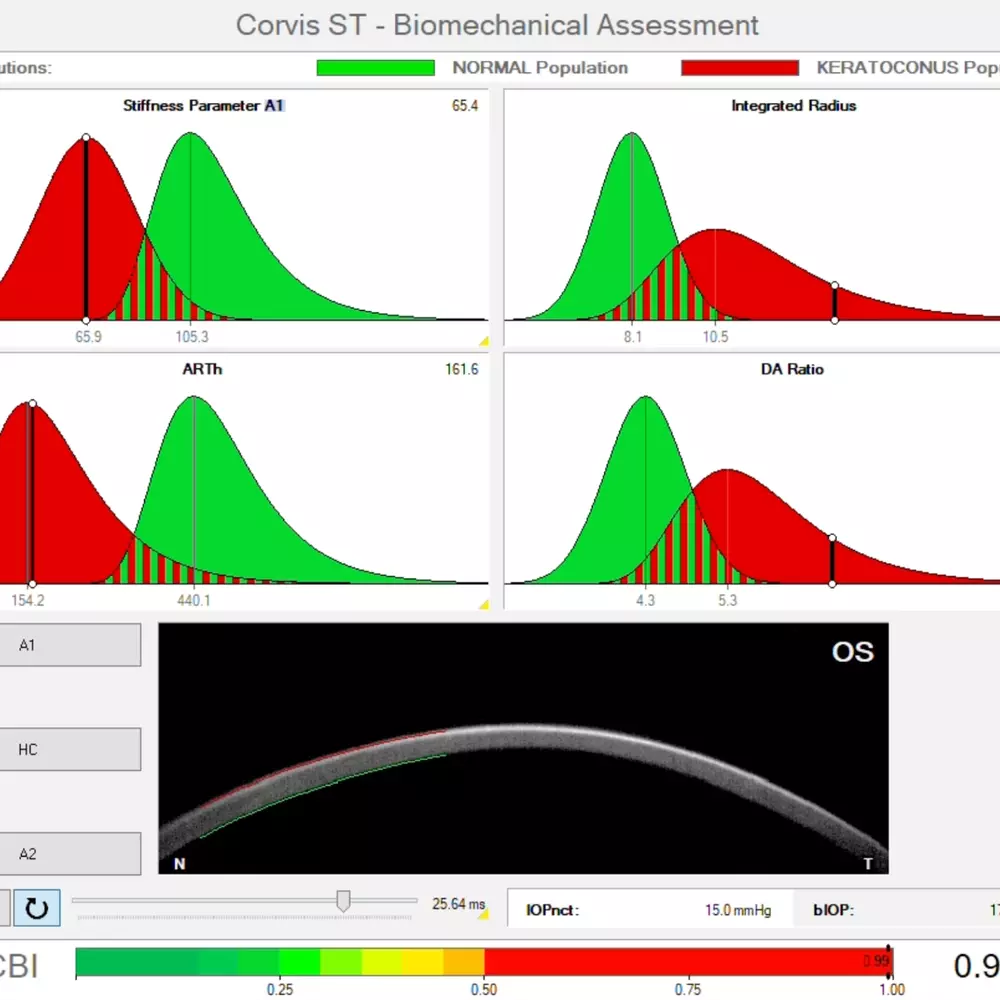
Eye with keratoconus
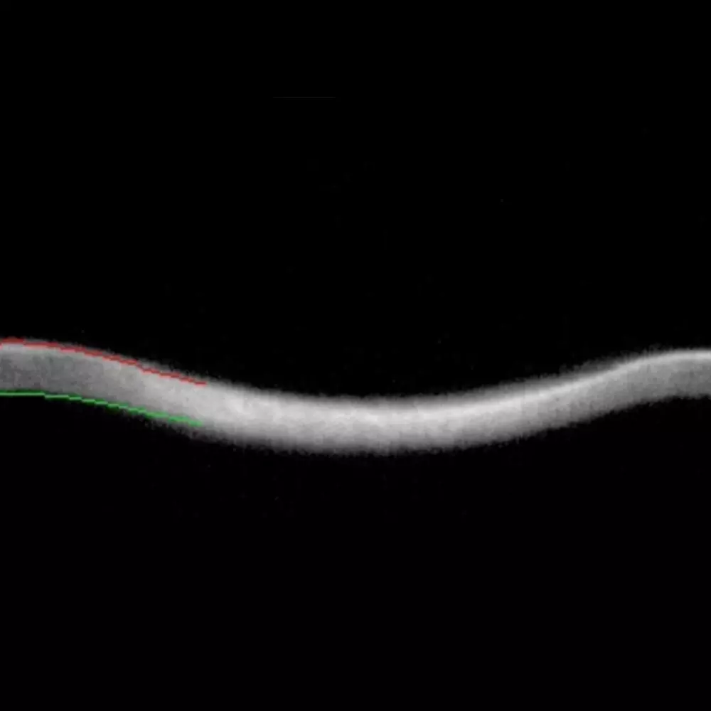
Eye with keratoconus
Schedule an Appointment for
Keratoconus Diagnosis
Please feel free to schedule an appointment for keratoconus screening. You can do so by phone, email, WhatsApp, or online via doctolib.de. We’d also be happy to call you back. We look forward to hearing from you.