
Refractive errors and eye conditions
What is Keratoconus?
Keratoconus is a progressive thinning and bulging of the cornea, which leads to high and irregular astigmatism. It is associated with fluctuating and declining visual acuity and, in advanced stages, cannot be corrected with glasses. The earlier it is detected and stabilized, the greater the chance of avoiding a corneal transplant.

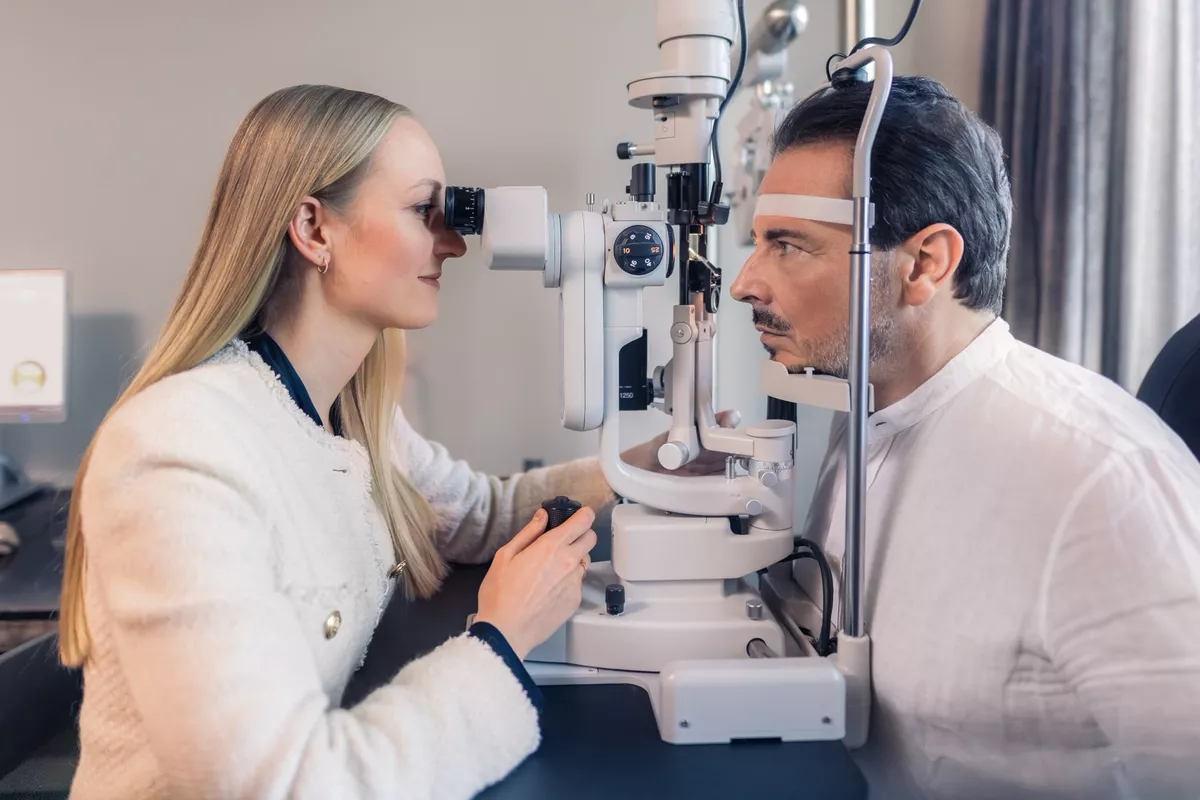
Unfortunately, the condition still goes undetected far too often. However, as part of our modern preliminary examinations—in which we assess the corneal structure not only with the Pentacam HR but also with state-of-the-art swept-source OCT devices—we are able to detect the typical corneal bulge and other characteristic features even in the early stages. We have been treating keratoconus since 2006 and recommend starting stabilizing therapy as early as possible.
Most of our keratoconus patients come to us initially hoping to live a life without glasses.
Schedule an appointment

Keratoconus is thought to be a disorder involving the collagen molecules in the connective tissue framework of the cornea, which leads to reduced collagen cross-linking and, consequently, reduced biomechanical stability of the cornea. The causes are complex and not yet fully understood.
The fact that it often runs in families suggests a genetic predisposition.
Immunological factors may also play a role.
However, the only thing that is certain so far is that vigorous and frequent rubbing of the eyes over many years—for example, in people with allergies—is a major risk factor for the development of keratoconus.
Thyroid dysfunction could also be a cause.
Studies show that UVA-riboflavin crosslinking appears to be effective in halting the progressive bulging of the cornea.
For this reason, we recommend that our keratoconus patients have their thyroid function tested so that, if necessary, they can begin treatment with thyroid hormones. The thyroid hormone thyroxine appears to have a significant influence on the biomechanics of the cornea: It increases the growth and stiffness of corneal tissue and affects the structure and properties of the collagen-protein complex.
As keratoconus progresses, the cornea becomes increasingly bulged and protrudes forward. A distinction is made between the “quiescent” form of keratoconus, known as “forme fruste,” and the advancing form, known as “progressive” keratoconus.
Symptoms
What are the symptoms of keratoconus?
Possible consequences of keratoconus include
increasing irregular astigmatism (corneal curvature),
unusual fluctuations and a decrease in visual acuity,
the perception of rings of light around light sources, known as “halos,”
as well as increased sensitivity to light and glare.
If the disease remains at a stage in which the full conical shape has not yet developed, it is referred to as “keratoconus forme fruste.” However, in about 20 percent of patients, progressive keratoconus develops, which advances rapidly. This can lead to tears in the posterior cornea, through which fluid from the anterior chamber enters the cornea, causing it to become cloudy. This is referred to as acute keratoconus. In the late stages, a corneal transplant is often the only effective treatment. Thanks to modern early detection and minimally invasive treatments, the condition does not have to progress to this point.
Shapes
Types of Keratoconus
Forme Fruste
In terms of symptoms, Forme Fruste is indistinguishable from normal astigmatism and is often discovered by chance during a thorough eye exam. It can be corrected with glasses, contact lenses, or even implantable contact lenses, and if the condition is stable, it does not require treatment but should be monitored and checked regularly. The "forme fruste" is ten times more common than the progressive form.
The progressive form of keratoconus
The progressive form of keratoconus is aggressive and often becomes apparent as early as the teenage years. The progressive bulging causes a curvature of the cornea, known as astigmatism. This irregularity of the cornea makes correction with glasses increasingly difficult. Contact lenses are a better option, as they apply pressure to the cornea and can smooth out the most severe irregularities. If the condition is not too advanced, lenses can provide good correction, even up to maximum visual acuity.
However, if the bulging of the cornea continues to increase, even hard contact lenses become less stable and may fall out. In addition, as the bulging progresses, the cornea becomes increasingly thinner and scarred below the center, which irreversibly impairs visual acuity.
UV riboflavin cross-linking is a promising treatment.
Studies show that UV riboflavin crosslinking can halt progressive corneal bulging because it stabilizes the tissue. Customized crosslinking can also improve vision. For this reason, Dr. Breyer and other keratoconus experts recommend undergoing this treatment as early as possible. Since the introduction of crosslinking, the number of corneal transplants required has been reduced by about half.
Keratoconus and Corneal Ectasia
Keratoconus is a specific form of corneal ectasia. Ectasia is a medical term. It refers to the bulging of a cavity. This type of corneal ectasia can also occur after LASIK surgery. It is believed that the flap incision weakens the cornea, leading to bulging of the cornea due to a lack of stability.
STEP Classification
New STEP Keratoconus Classification System
Optical coherence tomography (OCT) of the anterior segment provides higher image resolution than measurements taken with a rotating Scheimpflug camera. This allows the corneal epithelial layer to be distinguished from other layers with greater precision. In particular, the latest SS-OCT devices feature a light source with a longer wavelength, which achieves deeper tissue penetration. Mapping the epithelial layer thickness (ETM) and assessing changes in it have become established as important criteria for classifying keratoconus and for monitoring treatment. The new STEP classification system was developed based on this technology.2
The STEP system analyzes two key characteristics of the cornea to assess the stage of keratoconus:
ST value (Stroma Thinnest Thickness): the thinnest point of the middle layer of the cornea (stroma)—an important indicator of stability
EP value (Epithelial Standard Deviation): indicates the degree of variation in the thickness of the outer cell layer (epithelium)—a highly sensitive early warning sign of developing keratoconus
Stadiums in Berlin
Belin's Classification of Keratoconus
The ABCD classification system developed by Belin has now become the standard for staging keratoconus. It involves grading the following parameters:
A – anterior corneal curvature
, B – posterior corneal curvature
, C – pachymetry (corneal thickness) at the thinnest point, and
D – best-corrected spectacle visual acuity.
Regarding A and B: Both were measured in a 3.0-mm zone around the thinnest part of the cornea.
In the ABCD classification, there are 5 stages, ranging from 0 to 4, for all 4 parameters. A “−” is added if no scars are visible, a “+” for scars that allow the iris details to remain visible, and “++” for scars that obscure the iris details. This classification system makes it possible to document the disease’s progression over time, which clearly indicates whether the condition is stable or progressive.
-
A: Anterior surface curvature (3-mm zone) > 7.25 mm (< 46.5 dpt)
B: Rear surface curvature (3-mm zone) > 5.90 mm (< 57.25 dpt)
C: Thinnest pachymetry > 490 µm
D: Best-corrected spectacle visual acuity ≥ 20/20 (≥ 1.0)
Scarring: – -
A: Anterior surface curvature (3-mm zone) > 7.05 mm (< 48.0 dpt)
B: Rear surface curvature (3-mm zone) > 5.70 mm (< 59.25 dpt)
C: Thinnest pachymetry > 450 µm
D: Best-corrected spectacle visual acuity > 20/20 (< 1.0)
Scarring: −, +, ++ -
A: Front surface curvature (3-mm zone) > 6.35 mm (< 53.0 dpt)
B: Rear surface curvature (3-mm zone) > 5.15 mm (< 65.5 dpt)
C: Thinnest pachymetry > 400
µm D: Best-corrected spectacle visual acuity < 20/40 (< 0.5)
Scarring: −, +, ++ -
A: Front surface curvature (3-mm zone) > 6.15 mm (< 55.0 dpt)
B: Rear surface curvature (3-mm zone) > 4.95 mm (< 68.5 dpt)
C: Thinnest pachymetry > 300 µm
D: Best-corrected spectacle visual acuity < 20/100 (< 0.2)
Scarring: −, +, ++ -
A: Front surface curvature (3-mm zone) ≤ 6.15 mm (≥ 55.0 dpt)
B: Rear surface curvature (3-mm zone) ≤ 4.95 mm (≥ 68.5 dpt)
C: Thinnest pachymetry ≤ 300 µm
D: Best-corrected spectacle visual acuity < 20/400 (< 0.05)
Scarring: −, +, ++
Early Detection of Keratoconus
The thinning and bulging of the cornea caused by keratoconus can be detected at an early stage using modern diagnostic techniques. For the early detection of keratoconus, we rely on state-of-the-art OCT technology using the Anterion SS-OCT and Cirrus 6000 HD-OCT. Our specialist, Dr. Kristin L. Artz, is happy to assist you.

Treatment
Individualized Treatment of Keratoconus
Many factors play a role in deciding on the right treatment for keratoconus. According to the treatment protocol developed by an international group of experts, these include:
Your age
Your diagnosis
Your visual acuity with glasses or contact lenses
other factors, such as allergies that affect the eyes
An experienced ophthalmologist such as Dr. Detlev Breyer and our senior physician, Dr. Kristin Lena Artz, will be able to explain in more detail, following a consultation and a thorough examination, which treatment would be appropriate for you.
Our treatment approach is strictly guided by treatment safety and the principles of evidence-based medicine. Based on your diagnosis, we offer therapies that can slow the progression of the disease, as well as others that can improve your vision.
Early intervention protects visual acuity. Stabilization treatment should therefore be considered, especially in young patients.
Dr. Detlev R.H. Breyer

In this video on keratoconus treatment, host Nina Ruge asks Dr. Detlev Breyer which treatment is recommended for keratoconus. Dr. Breyer and his patient, Mr. Wilkes, describe UV riboflavin cross-linking (CXL) and iontophoresis cross-linking (iCXL)—one as a surgeon, the other as a patient—and explain the differences. The video was recorded in 2019. Today, we offer additional treatments to improve your vision.
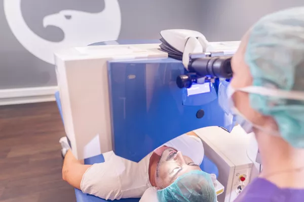



Implantable contact lenses are a good option for improving vision in cases of Forme Fruste keratoconus.
Dr. Detlev R.H. Breyer

Schedule an appointment
Do you have questions about keratoconus, or would you like to schedule an exam or consultation? If so, please feel free to schedule an appointment—by phone, email, WhatsApp, or online via doctolib.de. We’d also be happy to call you back. We look forward to hearing from you.